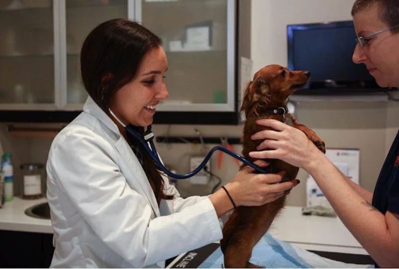
When a dog or cat is sick or in pain, the cause is not always visible from the outside. A pet may stop eating, begin limping, vomit repeatedly, develop changes in breathing, or simply act differently. A physical examination provides important clues, but sometimes a veterinarian needs to see what is happening inside the body.
Diagnostic imaging helps veterinarians examine internal structures without surgery. At Pure Paws Veterinary Care of Clinton Hill, imaging can play an important role in pet diagnostic care when symptoms or examination findings suggest that a closer look is needed. For pet parents, understanding why imaging is recommended can also make the diagnostic process less confusing.
What Is Veterinary Diagnostic Imaging?
Veterinary diagnostic imaging refers to technologies that create pictures of structures inside an animal’s body. Depending on the type of imaging, veterinarians can evaluate bones, joints, organs, soft tissues, the chest, and other areas.
Imaging does not automatically provide a diagnosis. Instead, veterinarians interpret the images alongside the pet’s symptoms, medical history, physical examination, and, when appropriate, bloodwork or other tests.
This combination helps narrow down possible causes and guides decisions about what should happen next.
How Does Imaging Help Veterinarians Find Health Problems?
Many medical conditions can produce similar symptoms. Vomiting, for example, may have several possible causes. Limping can result from problems involving bones, joints, muscles, or other tissues.
Imaging gives veterinarians additional information that cannot always be obtained through an external examination.
It may help a veterinarian:
- Identify fractures or changes involving bones and joints
- Examine the size, shape, and position of certain organs
- Look for abnormal fluid or gas
- Investigate possible intestinal obstruction
- Evaluate changes within the chest or abdomen
- Locate certain masses or abnormal tissue changes
- Assess some injuries following trauma
- Monitor known conditions over time
This is why imaging is an important part of pet diagnostic care rather than simply a test used for one particular disease.
What Can X-Rays Show in Dogs and Cats?
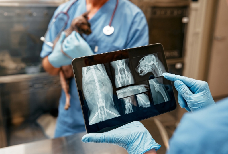
X-rays, also called radiographs, are among the most commonly used forms of veterinary imaging. They produce two-dimensional images based on how different tissues absorb X-rays.
Bones appear particularly clearly, making radiographs useful when veterinarians are concerned about fractures, joint changes, or certain skeletal abnormalities.
X-rays can also provide valuable information about the chest and abdomen. Depending on the clinical situation, they may help veterinarians evaluate the lungs, heart silhouette, gastrointestinal tract, bladder stones, swallowed objects, and other abnormalities.
However, an X-ray does not reveal every condition. If more detail is required, another imaging method or additional diagnostic testing may be recommended.
How Does Ultrasound Provide a Different View?
Ultrasound uses sound waves rather than X-rays to create images. It is particularly useful for examining many soft tissues and abdominal organs.
Unlike a standard radiograph, ultrasound produces images in real time. This allows a veterinarian to assess internal structures from different angles and observe certain movements.
An abdominal ultrasound may help evaluate organs such as the liver, kidneys, spleen, urinary bladder, and gastrointestinal tract. Ultrasound can also help identify some fluid accumulations, masses, changes in organ structure, and other abnormalities.
Because X-rays and ultrasound provide different types of information, one test does not necessarily replace the other. A veterinarian may recommend both when the findings from one study do not provide enough information.
Why Does a Veterinarian Choose One Imaging Test Over Another?
The appropriate imaging method depends on the pet’s symptoms, examination findings, and the body part to be evaluated.
For example, radiographs may be selected first when a veterinarian suspects a fracture or needs an overview of the chest. Ultrasound may provide more useful information when evaluating certain abdominal organs and soft tissues. More advanced tests, such as CT or MRI, can provide detailed cross-sectional images and may be recommended for particular conditions.
If your pet needs further evaluation, you may be advised to visit our veterinary hospital so the veterinarian can determine the appropriate diagnostic approach rather than choosing an imaging test based on symptoms alone.
Does My Pet Need Sedation for Diagnostic Imaging?
Not always. Many pets can undergo routine X-rays or ultrasound while awake. The decision depends on the imaging procedure, the pet’s comfort level, positioning requirements, and whether movement could interfere with image quality.
Some anxious or painful pets may need sedation. Advanced imaging procedures may also require sedation or anesthesia because the animal must remain very still.
Your veterinarian can explain what preparation is needed and why it is appropriate for your pet.
When Should Pet Owners Ask About Imaging?
Not every minor symptom requires diagnostic imaging. A brief, mild change that resolves quickly may be monitored under veterinary guidance. However, an examination should be considered when symptoms persist, recur, worsen, or interfere with normal eating, movement, breathing, or behavior.
Seek veterinary attention promptly for warning signs such as difficulty breathing, significant trauma, severe or worsening pain, repeated vomiting with weakness, a swollen abdomen with distress, inability to stand normally, or suspected ingestion of a foreign object or harmful substance.
These signs can have multiple causes. Imaging may be part of the evaluation, but only a veterinary examination can determine which tests are appropriate.
Imaging Can Help Turn Symptoms Into Useful Answers
When symptoms alone do not explain what is happening, imaging can provide another piece of the diagnostic picture. It allows veterinarians to examine internal structures, investigate possible causes, and determine whether further testing, monitoring, or treatment is needed.
For those seeking pet diagnostic care, an evaluation can help determine whether X-rays, ultrasound, or another diagnostic approach is appropriate. If you live in Brooklyn or a nearby community and are concerned about changes in your pet’s health, contact Pure Paws Veterinary Care of Clinton Hill. Our team, led by Dr. Slome, can evaluate your pet’s symptoms and determine whether diagnostic imaging may help provide the information needed to guide the next steps in care.
Frequently Asked Questions
1. What symptoms might lead a veterinarian to recommend imaging?
Persistent vomiting, limping, abdominal discomfort, breathing changes, unexplained pain, trauma, or changes in appetite may prompt further evaluation. These symptoms can have many causes, so a veterinarian will determine whether imaging is appropriate after examining the pet.
2. Can an X-ray detect every health problem in a pet?
No. X-rays provide valuable information about bones and certain changes in the chest or abdomen, but they cannot identify every condition. Ultrasound, laboratory testing, CT, MRI, or other tests may sometimes be needed for additional information.
3. Is veterinary ultrasound painful for dogs and cats?
Ultrasound is generally considered noninvasive and does not use ionizing radiation. Pets usually tolerate the procedure well, although their hair may need to be clipped, and some animals may require sedation if they are anxious, in pain, or unable to remain still.
4. How does a veterinarian decide between an X-ray and ultrasound?
The choice depends on the suspected problem and the structures being examined. X-rays are commonly used for bones and for broad views of the chest and abdomen, while ultrasound can provide more detailed, real-time views of many soft tissues and abdominal organs.
5. Does my pet need imaging after an injury?
It depends on the type and severity of the injury, as well as the findings during the examination. Imaging may be recommended after significant trauma to look for fractures or internal abnormalities that may not be apparent from the outside.
6. Can imaging find a health condition before symptoms become severe?
Imaging can sometimes identify structural changes that are not obvious during a physical examination. However, no imaging test detects every disease. Veterinarians use imaging, along with medical history, examination findings, and other diagnostic tests, to reach a clinical assessment.